Magnetic Resonance - Technology Information Portal Welcome to MRI Technology
Info
  Sheets

Out-
      side


 'cardiac imaging' 
SEARCH FOR    
 
  2 3 5 A B C D E F G H I J K L M N O P Q R S T U V W X Y Z
Result : Searchterm 'cardiac imaging' found in 0 term [] and 6 definitions [], (+ 17 Boolean[] results
previous     11 - 15 (of 23)     next
Result Pages : [1 2]  [3 4 5]
Searchterm 'cardiac imaging' was also found in the following services: 
spacer
News  (43)  Resources  (17)  Forum  (1)  
 
Echelon™ 1.5TInfoSheet: - Devices -
Intro, 
Types of Magnets, 
Overview, 
etc.
 
www.hitachimed.com/contentindex.asp?ID=971 From Hitachi Medical Systems America Inc.;
Hitachi expanded its portfolio with the Echelon™ 1.5T. The MRI scanner combines a compact magnet and a scalable 8-channel RF system with high-performance gradients and slew rate to select short echo times, small field of views, high matrices and thin slices. Standard features of the Echelon MRI system include higher-order active shim, RAPID (parallel imaging for use on brain MRI, body, cardiovascular imaging, and orthopedic coils), multiple coil ports, and an advanced reconstruction engine.
Device Information and Specification
CLINICAL APPLICATION
Whole body
CONFIGURATION
Short bore
Head, body coil, spine, breast, knee, shoulder, vascular multiple array coils.
SYNCHRONIZATION
Cardiac gating, ECG/peripheral, respiratory gating
PULSE SEQUENCES
SE, IR, FSE, FIR, GE, SG, BASG, PBSG, PCIR, DWI, Radial, Angiography: TOF, FLUTE (Fluoro-triggered bolus MRA), Time-resolved MRA
IMAGING MODES
Single, multislice, volume study
PIXEL INTENSITY
Level Range: -2,000 to +4,000
Sub millimeter
POWER REQUIREMENTS
208/220/240 V, single phase
CRYOGEN USE
Low cryogen boil-off
STRENGTH
30 mT/m
150 T/m/sec
Higher-order active shim
spacer
 
• Related Searches:
    • MRI Equipment
    • Superconducting Magnet
    • Excelart AG™ with Pianissimo
    • Hardware
    • MRI
 
Further Reading:
  Basics:
Echelon 1.5T
   by www.hitachimed.com    
MRI Resources 
Equipment - DICOM - Open Directory Project - Diffusion Weighted Imaging - PACS - Breast Implant
 
AIRIS II™InfoSheet: - Devices -
Intro, 
Types of Magnets, 
Overview, 
etc.MRI Resource Directory:
 - Devices -
 
www.hitachimed.com/products/airis_2.asp From Hitachi Medical Systems America Inc.;
the AIRIS II, an entry in the diagnostic category of open MR systems, was designed by Hitachi Medical Systems America Inc. (Twinsburg, OH, USA) and Hitachi Medical Corp. (Tokyo) and is manufactured by the Tokyo branch. A 0.3 T field-strength magnet and phased array coils deliver high image quality without the need for a tunnel-type high-field system, thereby significantly improving patient comfort not only for claustrophobic patients.
Device Information and Specification
CLINICAL APPLICATION
Whole body
CONFIGURATION
Vertical Field, Open MRI
QD Head, MA Head and Neck, QD C-Spine, MA or QD Shoulder, MA CTL Spine, QD Knee, Neck, QD TMJ, QD Breast, QD Flex Body (4 sizes), Small and Large Extrem., QD Wrist, MA Foot and Ankle (WIP), PVA (WIP)
SYNCHRONIZATION
Cardiac gating, ECG/peripheral, respiratory gating (2 modes)
PULSE SEQUENCES
SE, GE, GR, IR, FIR, STIR, FSE, ss-FSE, FLAIR, EPI -DWI, SE-EPI, ms - EPI, SSP, MTC, SARGE, RSSG, TRSG, MRCP, Angiography: CE, 2D/3D TOF
IMAGING MODES
Single, multislice, volume study
TR
SE: 30 - 10,000msec GE: 20 - 10,000msec IR: 50 - 16,700msec FSE: 200 - 16,7000msec
TE
SE : 10 - 250msec IR: 10 -250msec GE: 5 - 50 msec FSE: 15 - 2,000
SINGLE/MULTI SLICE
0.05 sec/image (256 x 256)
FOV
5cm to 42 cm continuous
2D: 2 - 100 mm; 3D: 0.5 - 5 mm
1280 x 1024
MEASURING MATRIX
512 x 512
PIXEL INTENSITY
Level Range: -2,000 to +4,000
Sub millimeter
MAGNET TYPE
Self-shielded, permanent
BORE DIAMETER
or W x H
110 x 43 cm
MAGNET WEIGHT
15,700 kg
H*W*D
79 x 111 x 73 cm
POWER REQUIREMENTS
208/220/240 V, single phase
COOLING SYSTEM TYPE
Air-cooled
STRENGTH
15 mT/m
2.0 m lateral, 2.5 m vert./long
Auto shimming, 3-axis/patient, and volume shim
spacer

• View the DATABASE results for 'AIRIS II™' (2).Open this link in a new window

MRI Resources 
Health - Corporations - Knee MRI - Homepages - Implant and Prosthesis pool - MRI Technician and Technologist Schools
 
Lantheus Medical Imaging, Inc.
 
www.radiopharm.com Lantheus Medical Imaging is a worldwide leader in medical imaging, with headquarters in North Billerica, Massachusetts, and nearly 700 employees worldwide with offices in Puerto Rico, Canada, and Australia.
The product portfolio includes ABLAVAR™ (gadofosveset trisodium) a blood pool contrast agent for magnetic resonance angiography MRA, Cardiolite® (Kit for the preparation of Technetium Tc99m Sestamibi), a cardiac perfusion imaging agent and DEFINITY® Vial for (Perflutren Lipid Microsphere) injectable suspension, the leading cardiac ultrasound contrast agent in the US.

MRI Contrast Agents:
Contact Information
MAIL
Lantheus Medical Imaging
Bldg. 200-2, 331 Treble Cove Rd.
N. Billerica, MA 01862
USA
PHONE
+1-800-362-2668
FAX
+1-978-436-7501
spacer

• View the DATABASE results for 'Lantheus Medical Imaging, Inc.' (3).Open this link in a new window


• View the NEWS results for 'Lantheus Medical Imaging, Inc.' (5).Open this link in a new window.
Searchterm 'cardiac imaging' was also found in the following services: 
spacer
News  (43)  Resources  (17)  Forum  (1)  
 
Virgo™InfoSheet: - Devices -
Intro, 
Types of Magnets, 
Overview, 
etc.MRI Resource Directory:
 - Devices -
 
www.canamglobal.com/mri/virgomri.html From Millennium Technology Inc. This open C-shaped MRI system eases patient comfort and technologist maneuverability. This low cost scanner is build for a wide range of applications. The Virgo™ patient table is detachable and moves on easy rolling castors. Able to accommodate patient weights up to 160 kg, the tabletop has a range of motion of 30 cm in the lateral direction and 90cm in the longitudinal direction. Images generated with this scanner can only be viewed (without data loss) on Millennium's proprietary viewing software.
Device Information and Specification
CLINICAL APPLICATION
Whole body
CONFIGURATION
C shaped
SYNCHRONIZATION
Standard cardiac gating, ECG/peripheral, respiratory gating
PULSE SEQUENCES
2D Spin Echo (single and multi-echo), 2D Inversion Recovery, 2D Sequential and 3D Volume Gradient Echo, 2D and 3D Spoiled Gradient Echo
IMAGING MODES
Localizer, single slice, multislice, volume, fast, POMP, multi slab, cine, slice and frequency zip, extended dynamic range, tailored RF
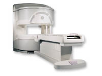
TR
steps of 1 msec
TE
steps of 1 msec
SINGLE/MULTI SLICE
Simultaneous scan and reconstruction;; 100 images/second reconstruction
400 mm
2D : 2 mm; 3D : 0.5 mm
MEASURING MATRIX
512x512
PIXEL INTENSITY
256 gray levels
MAGNET TYPE
Permanent
BORE DIAMETER
or Vertical Gap
44 cm
STRENGTH
15 mT/m
5-GAUSS FRINGE FIELD
3 m/3 m
Passive
spacer
MRI Resources 
Claustrophobia - Liver Imaging - Equipment - Safety Training - Sequences - Guidance
 
Signa OpenSpeed™InfoSheet: - Devices -
Intro, 
Types of Magnets, 
Overview, 
etc.MRI Resource Directory:
 - Devices -
 
www.gehealthcare.com/usen/mr/open_speed/index.html From GE Healthcare;
a friendly and less confining appearance targets the 7% of individuals who refuse to have an MRI because of claustrophobia. This open MRI system is also up to three times faster than other scanners, therefore the Signa OpenSpeed™ reducing exam time and scheduling issues. In addition, a swing table provides better access and supports up to 500 pounds.
Device Information and Specification
CLINICAL APPLICATION
Whole body
CONFIGURATION
Integrated transmit and receive body coil; 4 channel head array, 8 channel CTL array, 2 channel shoulder array, 3 channel large extremity array, 3 channel small extremity array, 4 channel foot array, 3 channel wrist array, 4 channel breast array
SYNCHRONIZATION
Standard cardiac gating, ECG/peripheral, respiratory gating
PULSE SEQUENCES
Standard: SE, IR, 2D/3D GRE and SPGR, Angiography: 2D/3D TOF, 2D/3D Phase Contrast;; 2D/3D FSE, 2D/3D FGRE and FSPGR, SSFP, FLAIR, EPI, optional: 2D/3D Fiesta, FGRET, Spiral, Tensor
IMAGING MODES
Localizer, single slice, multislice, volume, fast, POMP, multi slab, cine
TR
1.3 to 12000 msec in increments of 1 msec
TE
0.4 to 2000 msec in increments of 1 msec
SINGLE/MULTI SLICE
Simultaneous scan and reconstruction;; 100 images/second with Reflex 100
1cm to 40 cm continuous
2D: 0.8mm - 20mm 3D: 0.1mm - 20mm
1280 x 1024
MEASURING MATRIX
128x512 steps 32 phase//freq.
PIXEL INTENSITY
256 gray levels
0.08 mm; 0.02 mm optional
175 x 85 x 447 cm
MAGNET WEIGHT
8256 kg
H*W*D
530 x 175 x 250 cm
POWER REQUIREMENTS
200 - 480, 3-phase
COOLING SYSTEM TYPE
Liquid helium
0.03 L/hr, holds 300 L
STRENGTH
25 mT/m
5-GAUSS FRINGE FIELD
4.7 m/ 3.8 m
Computerized passive shimming during magnet setup, autoshim per series with automatic table motion to magnet isocenter for each prescription
spacer

• View the DATABASE results for 'Signa OpenSpeed™' (2).Open this link in a new window

 
Further Reading:
  News & More:
MR Surgical Suite, Improving surgical procedure quality (.pdf)
   by www3.gehealthcare.com    
MRI Resources 
MR Guided Interventions - Contrast Enhanced MRI - Quality Advice - MRI Technician and Technologist Career - Knee MRI - MRI Training Courses
 
previous      11 - 15 (of 23)     next
Result Pages : [1 2]  [3 4 5]
 Random Page
 
Share This Page
FacebookTwitterLinkedIn

MR-TIP    
Community   
User
Pass
Forgot your UserID/Password ?    


How AI will impact MRI :
only diagnostics 
saving time 
reducing cost 
makes planning obsolete 
reduce human knowledge 
not at all 

Look
      Ups


MR-TIP.com uses cookies! By browsing MR-TIP.com, you agree to our use of cookies.

Magnetic Resonance - Technology Information Portal
Member of SoftWays' Medical Imaging Group - MR-TIP • Radiology-TIP • Medical-Ultrasound-Imaging • 
Copyright © 2003 - 2024 SoftWays. All rights reserved. [ 1 May 2024]
Terms of Use | Privacy Policy | Advertising
 [last update: 2024-02-26 03:41:00]